OBSTETRICS CARE
Nurturing Beginnings with Advanced Obstetric Scans
We combine cutting-edge technology with compassionate care to redefine the journey of motherhood.
Our Obstetric Scans are designed to provide regular and comprehensive monitoring throughout your pregnancy, ensuring your health and well-being and that of your precious little one.

Prenatal Confirmation and Assessment (Viability Scan)
At 7-8 weeks, an ultrasound is conducted to check the healthof the pregnancy.
This is a simple and early confirmation of a viable pregnancy and allows for a precise determination of gestational age, and assessment of expected multiple pregnancies.
Risk Assessment at First Trimester
(Nuchal Translucency Scancombined with Blood Test)
Thorough assessment of the baby’s genetic risk through state-of-the-art imaging techniques combining with blood test. Performed at 11– 14 weeks of gestation, this is the opportunity to screen for common chromosomal abnormalities.
TYPES OF SCAN
At 7-8 weeks
A simple early confirmation of viability of pregnancy
Precise determination of gestational age
Assessment of expected multiple pregnancies
At 11-14 weeks
Confirm that the baby’s heartbeat is present
More accurate estimate of duration of pregnancy
Measure length of baby and assess structure of the fetus
(including measurement of fluid at the back of baby’s neck)
At 18-22 weeks
Can be recommended at 18 weeks also in event there are known risk factors for abnormalities
(or if parents want to know the sex of the baby at an earlier stage)Systematic check to assess that fetus’ structures appear as expected
Position of the placenta and blood flow through placenta is checked
At 28 weeks onwards
This is also called a ‘fetal growth scan’ or a ’wellbeing scan’. This is usually done in the third trimester after 28 weeks of gestation.
During this scan we measure the baby’s head, abdomen and limbs, and estimate the baby’s weight. This gives an idea about the growth of the baby. We also check the position of the baby in the womb, look at the baby’s movements, the amount of amniotic fluid, and the placental position.
We use Doppler ultrasound to check the blood flow in the umbilical cord and in the baby’s brain. In addition,we can also check to the blood flow the mother’s uterine arteries.
Usually between 22-24 weeks
Fetal echocardiography consists of the assessment of the baby’s heart before birth. Most children have a normal heart.
Approximately 8 in 1,000 babies born will show a heart defect. Half of these are major defects that can be identified before babies are born if a specialised scan of the fetal heart is performed. Although most complex abnormalities are well tolerated by the baby while in utero, treatment is usually required after birth and maybe as early as in the first few days of life, often due to changes in the way the blood goes round following delivery.
For the minority of babies that show an obvious major abnormality, the nature of the defect and available management options will be discussed with the parents and follow up organised as appropriate.
Minor abnormalities such as small holes in the heart may also be detected, but they do not disturb the way the heart works, even after birth. Treatment is usually unnecessary for minor abnormalities, but follow up will be arranged as appropriate. Apart from structural defects, some babies will need to be assessed for rhythm disturbances as well. These will be assessed and treatment options discussed at the time of the consultation.
Fetal echocardiography is usually carried out between 22-24 weeks of gestation and a single scan is generally adequate to assess the fetal heart. However, in some situations, the fetal heart can be assessed as early as 13-14 weeks of gestation and provide a lot of information and reassurance to some parents who are at risk of having a child with a congenital heart defect. In these cases, a second fetal echo is scheduled later on in pregnancy.
Recent studies have shown that the length of the neck of the womb (Cervical length) might indicate the chances for going into labour prematurely.
We will check this at the time of your 21 week scan or earlier if need be and then discuss the findings with you. In some women the doctor might want a cervical assessment earlier if there is any history to suggest miscarriages in the second trimester. This is specifically requested in women with the following history:
Prior history of preterm birth (<32 weeks gestation)
Previous cervical surgery (eg. cervical cone biopsy)
Women with a cervical suture
Women with suspected cervical incompetence
Multiple pregnancies
At our center, we understand your excitement and joy as your pregnancy progresses.
We offer you the opportunity to be reminded of this special time by keeping 3D/4D pictures and video clips of the scan of your unborn baby as they develop and grow.
We are able to get a 3D or 4D image when the baby is positioned.
Nice pictures and video clips are taken considering favourable position of the baby, maternal habitus and placenta location.
The scan is best done between 28-32 weeks.


RECENT BLOGS

Pregnancy and Bladder Control: Understanding and Managing Incontinence
Many expectant mothers experience bladder control issues during pregnancy. This guide offers insights on causes, symptoms, management strategies, and when to seek medical advice.
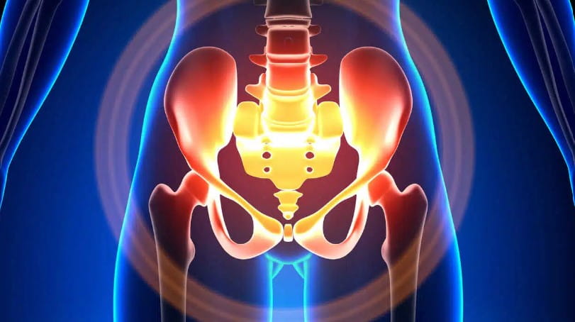
Pelvic Floor Dysfunction: Causes, Symptoms, and Treatments
Explore the comprehensive guide on pelvic floor dysfunction, including its causes, symptoms, diagnostic methods, and treatment options. Read real patient stories and understand the long-term benefits and risks of pelvic floor therapy.

Fetal Development: A Week-by-Week Guide for Expecting Mothers in Dubai
This blog outlines the week-by-week stages of fetal growth, from fertilization to birth. Learn how your baby develops during pregnancy and how to access prenatal care in Dubai.

Natural Birth vs. C-Section: An In-Depth Guide to Making the Best Choice for Your Delivery
Deciding between natural birth and C-section is a major choice for expectant mothers. This guide explores the pros and cons of each option, covering recovery, risks, and benefits to help you make an informed decision for your delivery.
